44 simple microscope diagram with labels
Microscope Drawing And Label at PaintingValley.com ... label microscope diagram compound parts light labeling functions microscopic blank labeled biology microscopy labelled beautiful Compound Microscope ... 496x600 35 0 Parts Of A Compound ... 500x469 27 0 Microscopic Drawing ... 1024x1024 21 4 Download The Diagram... 547x579 17 0 Microscope Labeling ... 270x350 17 0 Microscope Labeling ... Parts of a Simple Microscope - Labeled (with diagrams ... In this article, we are going to discuss the parts and functions of a simple microscope. image 1: The images above are all examples of a simple microscope. image source: laboratoryinfo.com. image 2: A simple microscope commonly used by students for studying minute objects. image source: imimg.com.
A Study of the Microscope and its Functions With a Labeled ... These labeled microscope diagrams and the functions of its various parts, attempt to simplify the microscope for you. However, as the saying goes, 'practice makes perfect', here is a blank compound microscope diagram and blank electron microscope diagram to label. Download the diagrams and practice labeling the different parts of these ...

Simple microscope diagram with labels
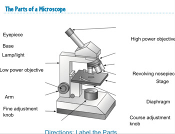
16 Parts of a Compound Microscope: Diagrams and Video ... Video: Parts of a compound Microscope with Diagram Explained. As a side note, the microscope used in this post is a great entry level or beginner microscope if you are trying to get someone interested in microscopes, microbiology, or science in general. PDF Basic Histo diagrams labelled in colour - 2005 These labelled diagrams should closely follow the current Science courses in histology, anatomy and ... created by Dr Carol Lazer during the period 2000-2005. STEREOLOGY: SLICING A 3-D OBJECT SIMPLE TUBE Do all microscope slides show 2-D slices of 3-D structures? No, slides can also be smears, where entire cells lie on the surface of the slide ... Label Microscope Diagram - EnchantedLearning.com Answers. Go to a microscope definition worksheet to print. EnchantedLearning.com. Label Microscope Diagram. Using the terms listed below, label the microscope diagram. Inventions and Inventors. arm - this attaches the eyepiece and body tube to the base. base - this supports the microscope.
Simple microscope diagram with labels. OptoHellas-Catalog-2020.pdf SPECULAR MICROSCOPE - A/B SCAN PACHYMETRY - TOPOGRAPHY ... The simple and easily operable SL-102 ... Morphology and density diagrams. Tiltable screen 10,4".24 σελίδες › topics › chemistryOptical Sensor - an overview | ScienceDirect Topics A far-field, epifluorescence microscope system for single tube spectroscopy was first proposed by Weisman and co-workers, and its schematic diagram is presented in Fig. 10.11. 23, 27 Visible images of sample morphology can be viewed through the eyepiece of the microscope or through a CCD camera. The samples are photo-excited by lasers, and ... Compound Microscope Parts - Labeled Diagram and their ... There are three major structural parts of a compound microscope. The head includes the upper part of the microscope, which houses the most critical optical components, and the eyepiece tube of the microscope. The base acts as the foundation of microscopes and houses the illuminator. The arm connects between the base and the head parts. quizlet.com › 290507793 › chapter-10-masteringbioChapter 10 MasteringBio Homework Flashcards - Quizlet Identify the membranes or compartments of the chloroplast by dragging the blue labels to the blue targets. Then, identify where the light reactions and Calvin cycle occur by dragging the pink labels to the pink targets. Note that only blue labels should be placed in blue targets, and only pink labels should be placed in pink targets.
Simple Microscope - Diagram (Parts labelled), Principle ... The working principle of a simple microscope is that when a lens is held close to the eye, a virtual, magnified and erect image of a specimen is formed at the least possible distance from which a human eye can discern objects clearly. Magnification formula. The magnification power of a simple microscope is expressed as: M = 1 + D/F. Where Compound Microscope Parts, Functions, and Labeled Diagram ... Compound Microscope Parts, Functions, and Labeled Diagram Parts of a Compound Microscope Each part of the compound microscope serves its own unique function, with each being important to the function of the scope as a whole. Simple Microscope - Parts, Functions, Diagram and Labelling Simple Microscope - Parts, Functions, Diagram and Labelling A microscope is one of the commonly used equipment in a laboratory setting. A microscope is an optical instrument used to magnify an image of a tiny object; objects that are not visible to the human eyes. Table of Contents The common types of microscopes are: What is a Simple microscope? › en › microscopeFluorescence Resonance Energy Transfer (FRET) Microscopy Presented in Figure 3 is a Jablonski diagram illustrating the coupled transitions involved between the donor emission and acceptor absorbance in fluorescence resonance energy transfer. Absorption and emission transitions are represented by straight vertical arrows (green and red, respectively), while vibrational relaxation is indicated by wavy ...
PDF Parts of a Microscope Printables - Homeschool Creations and 40x. The eyepiece on a microscope magnifies at 10x, so when used together, the 4x lens magnifies an item 40x, the 10x magnifies 100x, and the 40x magnifies 400x. (note: for typical student microscope -other microscopes will vary) •Which part of the microscope rotates so another person can look through the eyepiece › books › NBK21116Mapping Genomes - Genomes - NCBI Bookshelf Draw a diagram showing how a double-stranded cDNA is synthesized. 15. Define the term ‘mapping reagent’ and explain how a panel of radiation hybrids is used as a mapping reagent. 16. Explain how a clone library is used as a mapping reagent. 17. Draw a diagram to show how a sample of a single human chromosome can be obtained by flow cytometry. en.wikipedia.org › wiki › FluorescenceFluorescence - Wikipedia Fluorescence is the emission of light by a substance that has absorbed light or other electromagnetic radiation.It is a form of luminescence.In most cases, the emitted light has a longer wavelength, and therefore a lower photon energy, than the absorbed radiation. Compound Microscope- Definition, Labeled Diagram ... A standard Microscope has three to four Objective Lenses which range from 4X to 100X. Stage Clips are metal clips that held the slide in place. Arm and Base The Arm connects the Body Tube to the base of the Microscope. The Base supports the Microscope and its where Illuminator. Illuminator and Stage
Microscope Diagram Labeled, Unlabeled and Blank | Parts of a Microscope | Science printables ...
› cells › bactcellInteractive Bacteria Cell Model - CELLS alive Ribosomes: Ribosomes give the cytoplasm of bacteria a granular appearance in electron micrographs.Though smaller than the ribosomes in eukaryotic cells, these inclusions have a similar function in translating the genetic message in messenger RNA into the production of peptide sequences (proteins).

Diagram Of The Microscope - ClipArt Best
Microscope Labeling - The Biology Corner 1) Start with scanning (the shortest objective) and only use the COARSE knob . Once it is focused… 2) Switch to low power (medium) and only use the COARSE knob . You may need to recenter your slide. Once it is focused.. 3) Switch to high power (long objective).

All Saints Online: Diagram for Labelling: Microscope
Labeling Microscope Worksheet | Teaching Resources docx, 300.56 KB. A straightforward worksheet in which students are required to identify the parts of a basic microscope. Tes classic free licence.

The Microscope: Create a Labelled Diagram | Teaching Resources
A Study of the Microscope and its Functions With a Labeled ... sciencestruck.com A Study of the Microscope and its Functions With a Labeled Diagram To better understand the structure and function of a microscope, we need to take a look at the labeled microscope diagrams of the compound and electron microscope. These diagrams clearly explain the functioning of the microscopes along with their respective parts.

White Blood Cells Diagram
Parts of the Microscope with Labeling (also Free Printouts ... Parts of the Microscope with Labeling (also Free Printouts) A microscope is one of the invaluable tools in the laboratory setting. It is used to observe things that cannot be seen by the naked eye. Table of Contents 1. Eyepiece 2. Body tube/Head 3. Turret/Nose piece 4. Objective lenses 5. Knobs (fine and coarse) 6. Stage and stage clips 7. Aperture

# 58 The immune system - Phagocytes | Biology Notes for A level
Microscope labeled diagram - SlideShare Microscope labeled diagram 1. The Microscope Image courtesy of: Microscopehelp.com Basic rules to using the microscope 1. You should always carry a microscope with two hands, one on the arm and the other under the base. 2. You should always start on the lowest power objective lens and should always leave the microscope on the low power lens when you finish using it. 3.

anatomyforme: 2008-04-06
Label the microscope - Science Learning Hub All microscopes share features in common. In this interactive, you can label the different parts of a microscope. Use this with the Microscope parts activity to help students identify and label the main parts of a microscope and then describe their functions. Drag and drop the text labels onto the microscope diagram. If you want to redo an answer, click on the box and the answer will go back to the top so you can move it to another box.

A typical animal cell (as seen in an electron microscope) Medical Ima…
quizlet.com › 535464645 › masteringbiology-ch-10MasteringBiology: Ch 10 Flashcards & Practice Test | Quizlet Drag the labels onto the flowchart to show the relationship between the production of photons by the sun (Engelmann's light source) and the distribution of bacteria that Engelmann observed under his microscope. Not all labels will be used.

Microscope Labeling Worksheet 42 New Microscope Labeling Thinglink Worksheets Microscopes ...
Simple Microscope Definition, Magnification, Parts And Uses Aim: To make a simple microscope with the help of water. Apparatus Required A glass of water Fuse wire Object to view (newspaper works well due to its fine print) Procedure Make a loop of the fuse wire around 2 mm wide. Dip it in water so that a drop is made in the loop. Hold it near to your eye and take a close look at the object you have chosen.
All Saints Online: Diagram for Labelling: Microscope
Microscope Parts and Functions With Labeled Diagram and ... The specimen is placed on the glass and a cover slip is placed over the specimen. This allows the slide to be easily inserted or removed from the microscope. It also allows the specimen to be labeled, transported, and stored without damage. Stage: The flat platform where the slide is placed.

Microscope Clip Art at Clker.com - vector clip art online, royalty free & public domain
Microscope Labeling - The Biology Corner Students label the parts of the microscope in this photo of a basic laboratory light microscope. Can be used for practice or as a quiz. Name_____ Microscope Labeling . Microscope Use: 15. When focusing a specimen, you should always start with the _____ objective.
Simple columnar epithelium
Parts of a microscope with functions and labeled diagram Structural parts of a microscope and their functions Figure created with biorender.com Figure: Diagram of parts of a microscope There are three structural parts of the microscope i.e. head, base, and arm. Head - This is also known as the body. It carries the optical parts in the upper part of the microscope. Base - It acts as microscopes support.
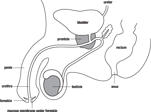
Male Reproductive System | Free Images at Clker.com - vector clip art online, royalty free ...
Compound Microscope: Definition, Diagram, Parts, Uses ... Compound microscope is a type of optical microscope that is used for obtaining a high-resolution image. There are more than two lenses in a compound microscope. Learn about the working principle, parts and uses of a compound microscope along with a labeled diagram here.

All Saints Online: Microscope Part Functions
Simple Microscope - Definition, Types, Working Principle ... Working of Simple Microscope. A simple microscope consists of a convex lens of a short focal length. The below figure shows the ray diagram which subsequently forms the image of an object (or we can say a source of light). (Image will be Updated soon) F is the focal length of the lens.
Simple Microscope Labeled Diagram - Micropedia
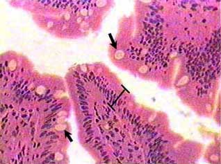
Simple Columnar Epithelium Labeled Diagram Simple Columnar Epithelium: A Labeled Diagram and Functions Epithelium is a tissue that lines the internal surface of the body, as well as the internal organs. Simple epithelium is one of the types of epithelium that is divided into simple columnar epithelium, simple squamous epithelium, and simple cuboidal epithelium.

The Microscope: Create a Labelled Diagram | Teaching Resources
Microscope Types (with labeled diagrams) and Functions A compound microscope: Is used to view samples that are not visible to the naked eye. Uses two types of lenses - Objective and ocular lenses. Has a higher level of magnification - Typically up to 2000x. Is used in hospitals and forensic labs by scientists, biologists and researchers to study micro organisms. Compound microscope labeled diagram.

*Simple Squamous Epithelium*.........................(LOCATION: capillary walls, alveoli of ...
Microscope, Microscope Parts, Labeled Diagram, and Functions Revolving Nosepiece or Turret: Turret is the part of the microscope that holds two or multiple objective lenses and helps to rotate objective lenses and also helps to easily change power. Objective Lenses: Three are 3 or 4 objective lenses on a microscope. The objective lenses almost always consist of 4x, 10x, 40x and 100x powers. The most common eyepiece lens is 10x and when it coupled with ...
Post a Comment for "44 simple microscope diagram with labels"